
Mystery of Knee Ligament Changes After Injury and Their Role in Arthritis
Authors: Ramos-Mucci, L., Elsheikh, A., Keenan, C., Eliasy, A., D’Aout, K., Bou-Gharios, G., Comerford, E., Poulet, B.
Journal: Arthritis Research & Therapy
Publication Date: May 2022
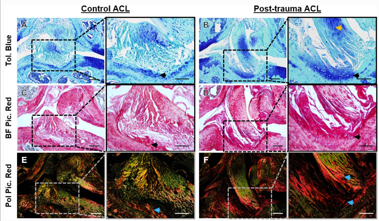
Representative histological staining in the anterior cruciate ligament (ACL) of control and post-trauma mouse knee joints. A, B Toluidine blue (Tol. Blue) staining of healthy and post-trauma ACL showed Tol. Blue staining in the mid-substance and tibial enthesis of the post-trauma ACL (black arrow). C, D Brightfield Picrosirius Red (BF Pic. Red) staining confirmed rounded-cell morphology in the tibial enthesis of the post-trauma ACL (black arrow). E, F Polarised Picrosirius Red (Pol Pic. Red) showed red collagen birefringence in the tibial enthesis extending towards the mid-substance of the post-trauma ACL (blue arrows). Scale is 100 μm and 50 μm for lower and higher magnification. Post-trauma ACL images are from + 4-weeks post-trauma
Summary:
Knee injuries, especially those involving ligaments, are common among athletes and can lead to a painful condition called post-traumatic osteoarthritis (PTOA). Despite their importance in stabilising the knee joint, the role of ligaments in PTOA development is not well understood. Our research aimed to investigate the structural and mechanical changes that occur in a key knee ligament, the anterior cruciate ligament (ACL), after a knee injury.
Using a mouse model of knee trauma, we discovered that injured knee joints showed increased joint space mineralisation, indicating the progression of osteoarthritis. The ACLs in injured knees exhibited changes in their extracellular matrix, including alterations in collagen and proteoglycan composition. Additionally, the injured ACLs showed decreased stiffness and altered viscoelastic properties, which are essential for proper ligament function.
Our findings reveal that changes in the structure and function of the ACL after injury can compromise the health of the knee joint and play a role in the development of PTOA. Understanding these changes can help develop new treatment strategies to improve ligament function and potentially prevent PTOA progression in the future.