
Corneal Disease Detection with Multi-Meridian Imaging
Authors: Curatolo, A., Birkenfeld, J.S., Martinez-Enriquez, E., Germann, J.A., Muralidharan, G., Palací, J., Pascual, D., Eliasy, A., Abass, A., Solarski, J., and Karnowski, K.
Journal: Biomedical Optics Express
Publication Date: Oct 2020
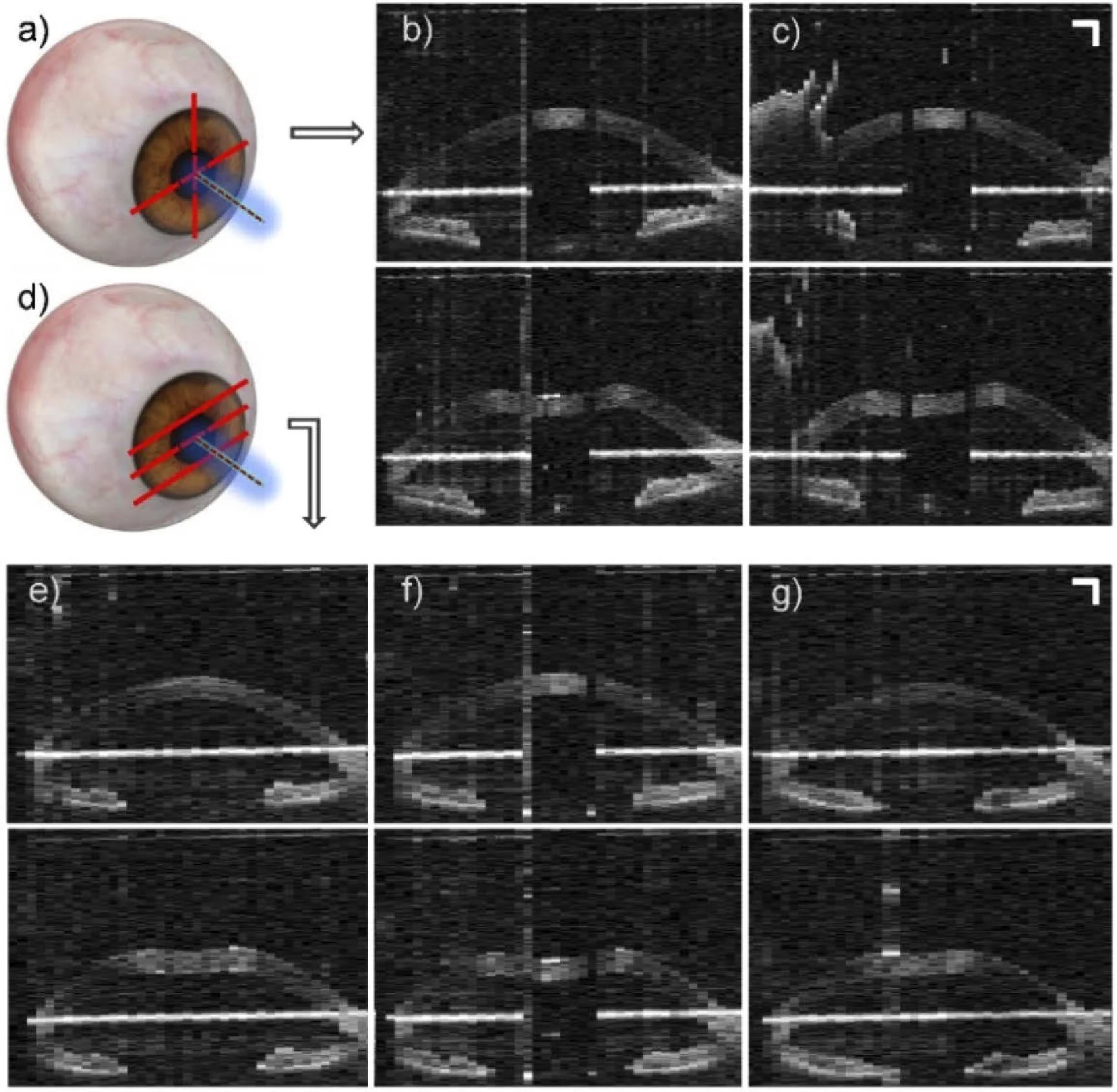
(a) “Cross-meridian” scan pattern schematic. OCT in vivo human corneal deformation frames before air-puff (top) and at maximum deformation (bottom), for (b) horizontal meridian and (c) vertical meridian. (d) “Three horizontal planes” scan pattern schematic. OCT in vivo human corneal deformation frames before air-puff (top) and at maximum deformation (bottom), for (f) the horizontal meridian, (e) 2 mm above, and (g) 2 mm below. Scale bars: 1 mm
Summary:
Corneal biomechanics play a crucial role in maintaining healthy eyes and good vision. The detection of corneal diseases such as keratoconus, a progressive disorder causing the cornea to thin and bulge, can be challenging. Early detection is essential for treating this disease before significant corneal changes occur. Current commercial systems are limited in their ability to monitor corneal deformation, capturing information only on one corneal meridian.
Our research introduces a custom-developed swept-source optical coherence tomography (SSOCT) system, combined with a collinear air-puff excitation. This new technology allows us to acquire dynamic corneal deformation on multiple meridians, improving the detection of biomechanical abnormalities in the cornea.
Our study involved testing the multi-meridian corneal deformation profiles on healthy human eyes, porcine eyes with varying controlled intraocular pressure, and a keratoconus-mimicking porcine eye. We were able to detect deformation asymmetries otherwise missed on a single meridian, which will significantly aid in corneal biomechanics diagnostics and pathology screening.
In conclusion, our novel multi-meridian corneal deformation imaging system offers a substantial advancement in detecting corneal diseases, providing crucial information for early intervention and improved patient outcomes.