
The Magic of Ortho-K Lenses: A Closer Look at Corneal Changes
Authors: Ran, Z., Moore, J., Jiang, F., Guo, H., Eliasy, A., Lopes, B.T., Bao, F., Jiang, J., Abass, A., Elsheikh, A.
Journal: Royal Society Open Science
Publication Date: Dec 2021
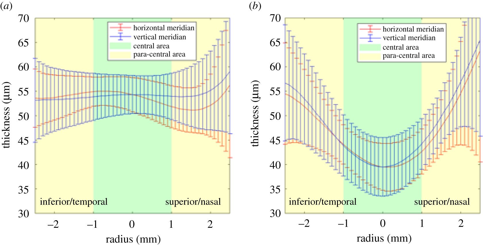
Mean epithelial thickness (a) before and (b) after Ortho-K lens wear.
Summary:
Orthokeratology (Ortho-K) lenses have been a game-changer for people with myopia, offering a non-surgical way to reshape the cornea and improve vision. Our research sought to understand the changes in corneal thickness and curvature after wearing Ortho-K lenses. We used a new, custom-built method to automatically detect corneal surfaces in optical coherence tomography (OCT) scans, allowing for precise measurements of changes in the cornea.
After one month of uninterrupted overnight wear of Ortho-K lenses, we found that the central corneal epithelium, the thin outer layer of the cornea, thinned by 23.8% on average. The anterior corneal surface became flatter, while the anterior and posterior surfaces of the stroma, the thicker middle layer, remained largely unchanged. The reduction in myopic refractive error was mainly due to changes in the corneal epithelium thickness profile.
Our study introduces a new method for automatically detecting corneal surfaces in OCT scans and sheds light on the mechanisms behind the effectiveness of Ortho-K lenses. These findings contribute to a better understanding of how Ortho-K lenses work, which may lead to further improvements in the design and use of these lenses for myopia treatment.